

Common Liver Lesions
 |
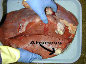 |
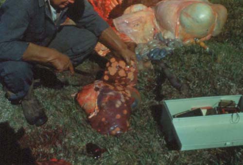
| Both of the above pictures are examples of liver abscesses. The abscesses are the yellow to white spots on the liver. Not all abscesses may be evident from the surface of the organ. It is important to cut into the organ to see if there is abnormal tissue or abscesses within the organ. | |
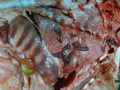
| These are examples of liver flukes. These travel through the liver causing damage to the liver. You may be able to see their tracts. These are usually seen in marshy areas that have snails. | 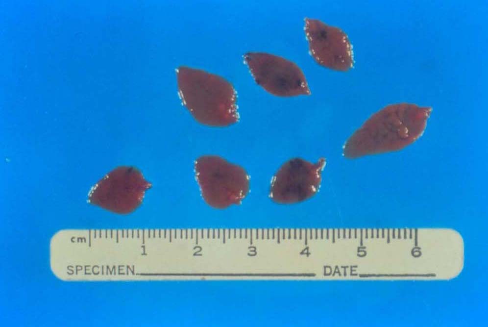 |
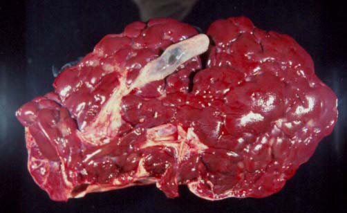 |
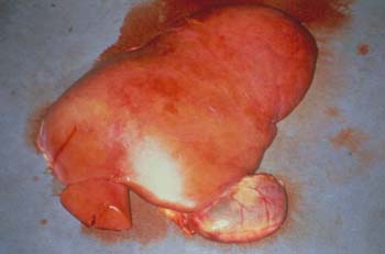
Notice how the liver looks lobated like a kidney. This is due to scaring or fibrosis from a chronic disease. |
| This liver has a "nutmeg" appearance. White areas surrounded by red areas. This may be seen in congested livers associated with heart failure. | 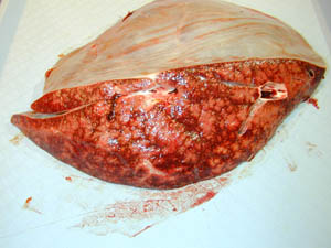 |
 |
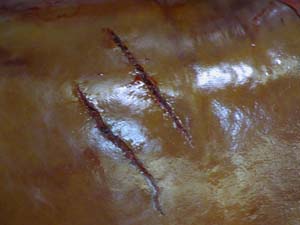 |
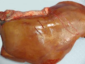
| This is an example of a fractured liver. These cuts in the liver where not created by a knife, but instead where caused by blunt trauma to the liver. Knife cuts will be smooth where these are jagged. The photo to the right is a close-up of the left photo. | |
 |
Note that this liver has a stripped appearance. This is an insignificant finding and is due to the animal laying on its side after death. The pressure against the rib cage forces the blood out and will cause a stripped appearance such as this. |
 |
This photo depicts a fatty liver. These livers will appear pale, have rounded edges, and may float in water. |